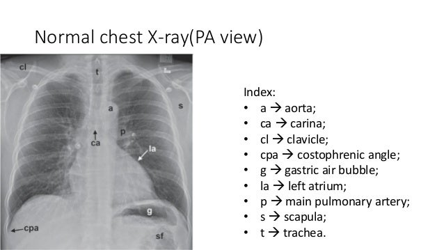
Pleural effusion volume mL distance cm x 90. For pleural effusion of less than 50 ml the x-ray has to be taken in the lying position with the patient turned towards the side of effusion.

Pleural Effusion Radiology Reference Article Radiopaedia Org
When air enters into the pleural 000030 space it will rise to the most superior part of the thorax.

How to estimate pleural effusion on x ray. A chest radiograph prediction rule. This is a chest X Ray PA view showing homogenous opacity involving the whole left lung field with deviation of the trache and mediastinum to the right sideT. The amount of pleural fluid volume can be estimated with the simplified formula.
The volume of a pleural effusion can be estimated from the chest radiograph appearance with a reasonable degree of accuracy. A prediction rule was devised for estimating pleural effusion volume on the basis of the presence or absence of a meniscus on chest radiographs. The most dependent recess of the pleura is the posterior costophrenic angle.
The rule was tested and validated using separate data sets obtained from a retrospective review of patients having both a chest radio- graph and computed tomography CT scan the gold standard within. The error rate in size estimation was 41 53129 for. The mortality rates associated with Pleural E usion seems to be dependent on a variety of factors however the initial diagnosis of this condition is key for.
We provide guidelines for estimating PF volumes on upright frontal and lateral CXRs. Asymmetric pleural effusions. Eventually a meniscus will be seen on frontal films seen laterally and gently sloping medially note.
Right and left pleural effusion. This video shows the differential diagnosis of pleural_effusion. Pleural effusions caused by heart failure may not be symmetrical.
Fluid gathers in the lowest part of the chest according to the patients position. Based on the curve and its trend the more reasonable cutoff would be set at 50 of the hemithorax for small moderate and large respectively. The fluid may be transude exudate blood chyle or rarely bile.
What is abnormal in this chest xray. In addition it was consistent with the broadly accepted classification on the chest radiograph in. The effusion is considered to be an exudate when the pleural fluid protein level divided by serum protein level is greater than 05 when pleural fluid LDH divided by serum LDH is greater than 06 and when pleural fluid LDH is more than two-thirds the upper limit of normal serum LDH.
We also confirm that the lateral radiograph is more sensitive for detection of small pleural effusions with blunting of the posterior CPA only correlating with a mean of 26 mL of PF. A pleural effusion is a collection of fluid in the pleural space. Blunting of the cardiophrenic angle.
Mean prediction error of V using Sep was 1584-1606 ml. Fluid has accumulated in the right pleural space the right costophrenic angle is not visible. In order to identify pneumothorax we need to identify the black air within the pleural space and to differentiate that from the air within the lungs.
Chest x-ray is a simple test to diagnose pleural effusion see figures 145 and 6. US pleural effusion size estimation correlated most closely with actual volume of fluid drained r 0833 N 179 P 00001 vs. Pleural fluid casts a shadow of the density of water on the chest radiograph.
Miscellaneous such as pacemakers catheters etc. Pleural fluid volume estimation. Blunting of the costophrenic angle.
The second Goecke formula measures the distance between the lung base and the mid-diaphragm the subpulmonary height height of the dome of the diaphragm is connected to the lung base the line being perpendicular to. Pleural effusion is the accumulation of fluid in the pleural space ie. This patient does not have one of the following diseases.
Infection heart failure cancer inflammatory conditions such as lupus cirrhosis post heart surgery pulmonary embolism clots to the lungs amongst other causes. If the patient is upright when the X-ray is taken then fluid will surround the lung base forming a meniscus a concave line obscuring the costophrenic angle and part or all of the hemidiaphragm. An example of frontal x-ray with Pleural E usion is shown in Figure 2.
The amount of fluid to be evident on a posteroanterior film is 200 mL whereas costophrenic angle blunting can be appreciated on a lateral film when approximately 50 mL of fluid has accumulated. Standard posteroanterior and lateral chest radiography remains the most important technique for initial diagnosis of pleural effusion. Chest X Ray differential diagnosis for pleural effusion - YouTube.
A new simple method for estimating pleural effusion size on CT scans. This is a common finding on chest X-ray which can have many causes such as. It also shows how to differentiate it fromdiaphragmatic.
How does a chest X-ray diagnose pleural effusion. This patient with heart failure had been nursed lying on their right side before this X-ray was taken. Easy quantification of pleural fluid may help to decide about performing thoracentesis in high-risk patients although thoracentesis under ultrasound guidance appears to be a safe procedure.
This position is called lateral decubitus position. This formula-based method of. X-rays from e usions with over 200ml of uid or 50ml in some lateral x-rays10.
A pleural effusion is the accumulation of fluid between the layers of pleura that cover the lung. If further air accumulates then the air will accumulate lateral and even inferior to the lung. Principles of Reading 1.
V ml20 x Sep mm. PLEURAL EFFUSION PERICARDIAL EFFUSION. No effusion is present in the left pleural space.
Pleural effusion lateral decubitus view A lateral decubitus chest radiograph with the side containing the pleural effusion placed down dependent demonstrate smaller amounts of free-flowing pleural effusions 1 millimeter of thickness of pleural fluid in. Use of the formula d2 x l readily enables estimation of pleural effusion volume from CT from two simple measurements. Between the visceral and parietal layers of pleura.
Fluid within the horizontal or oblique fissures. CXR r 0548 N 129 P 0001 and CT r 0489 N 107 P 0001.

Pdf Smart Pleural Effusion Drainage Monitoring System Establishment For Rapid Effusion Volume Estimation And Safety Confirmation Semantic Scholar

Estimate Pleural Effusion Volume Youtube

Pleural Effusion X Ray Findings

Serial Chest X Rays Cxrs Of Patient 1 A First Presentation With Download Scientific Diagram

Tidak ada komentar:
Posting Komentar